

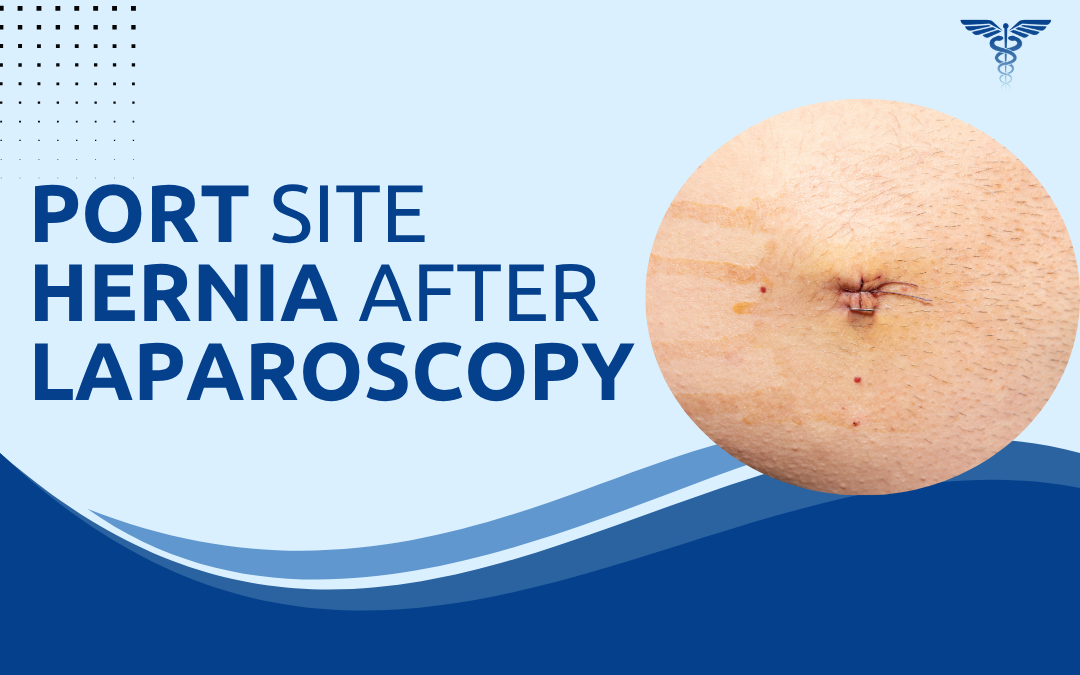
Port site hernias occur when abdominal tissue or bowel pushes through a weak spot at a laparoscopic incision site. The wound may look healed on the outside, but if the fascia underneath wasn’t closed properly, a gap remains.Most patients don’t expect this after “keyhole” surgery the incisions are small, so it seems low-risk. But incision size doesn’t equal defect risk. Even a 10–12 mm port site can leave a real fascial gap, and a hernia can develop there weeks, months, or years later.
According to Dr. Rajeev Premnath, a trusted Laparoscopic Surgeon in Bangalore, “Port site hernias are one of the more underrecognised complications of laparoscopic surgery. Patients assume small incisions mean no hernia risk. The skin heals quickly, so everything appears fine. But the fascial defect underneath is what determines whether a hernia forms, and that layer requires deliberate closure at port sites of 10 mm and above.”
Why Does a Port Site Hernia Happen?
The mechanism is the same as any other hernia. Raised intra-abdominal pressure pushes tissue through a defect in the abdominal wall. At a port site, that defect is the incision made to insert a trocar during laparoscopic surgery. Several factors increase the risk:
- Port size: Incisions of 10 mm and above carry the highest risk. The fascia at these sites must be formally closed. Smaller 5 mm ports rarely herniate unless wound infection occurs.
- Inadequate fascial closure: The skin closing over a port site does not close the deeper fascial layer. If only the skin is sutured, the abdominal wall defect remains open underneath.
- Wound infection: Infection breaks down the healing tissue, widens the defect, and significantly raises the likelihood of herniation. This is one of the most preventable contributing factors.
- Patient factors: Obesity, chronic cough, constipation, and heavy lifting in the early post-operative period all raise intra-abdominal pressure and stress the healing wound. The relationship between obesity and hernia surgery complications is well established.
- Trocar site location: Umbilical port sites herniate more often than lateral sites because the umbilicus is a natural weak point in the abdominal wall, similar to why umbilical hernias in adults occur at the same location.
- Type of procedure: Longer or more complex procedures with wider instrument manipulation carry a slightly higher rate of port site herniation compared to shorter straightforward operations.
How Is a Port Site Hernia Treated?
Surgery is the only definitive treatment. Port site hernias do not resolve on their own. Watchful waiting is appropriate only for very small, asymptomatic defects in patients unfit for surgery. The surgical approach depends on defect size and complexity. Patients with additional risk factors may require high risk hernia surgery assessment before proceeding.
-
- Laparoscopic repair: The defect is identified under camera visualisation, hernial contents are reduced, and mesh is placed intraperitoneally with adequate overlap. This mirrors the technique used for other laparoscopic hernia repairs and carries a low recurrence rate. Most patients are discharged the same day, consistent with a day care surgical approach.
- Open repair: Used for larger defects, recurrent hernias, or cases where bowel resection is required. A direct incision is made over the defect, contents are reduced, and mesh is placed in the appropriate plane.
- Mesh reinforcement: Mesh is standard in most port site hernia repairs. Primary suture closure alone carries a higher recurrence rate, particularly for defects above 1 cm. The principles are the same as those governing hernia mesh repair generally.
- Recovery: Days 1 to 7 involve light activity only. Return to desk work by weeks 2 to 4. Full activity including exercise for most patients by weeks 4 to 6 after laparoscopic repair. Open repair recovery extends to 6 to 8 weeks. The full hernia surgery recovery timeline applies here.
Why Choose Dr. Rajeev Premnath?
Dr. Rajeev Premnath is a General and Laparoscopic Surgeon with MBBS, MS (Gen Surg.), FRCS (Glasg.), FEBS, FICS, FACS, FIAGES, FMAS, and a Diploma in Laparoscopy from France. He serves as Head of Day Care Surgery at Ramakrishna Group of Hospitals and has over 20 years of experience in laparoscopic procedures across all complexity levels.
Port site hernia repairs here are assessed individually. The approach, mesh selection, and discharge plan are tailored to the defect size, patient fitness, and the clinical picture at presentation. High-risk patients receive a thorough pre-operative evaluation before any operative decision is confirmed.
Noticed a new bulge near a previous laparoscopic scar?
FAQs
What is a port site hernia?
A port site hernia is a hernia that forms at one of the small incision sites made during laparoscopic surgery. It occurs when the fascial layer beneath the skin is not adequately closed, leaving a defect through which abdominal tissue or bowel can protrude.
How long after laparoscopic surgery can a port site hernia develop?
A port site hernia can develop weeks, months, or even years after laparoscopic surgery. Early hernias typically result from inadequate fascial closure. Late hernias often develop gradually as intra-abdominal pressure widens a partial defect over time.
Is port site hernia repair done laparoscopically?
Yes, most port site hernia repairs are performed laparoscopically. The defect is identified under camera vision, contents are reduced, and mesh is placed intraperitoneally. Open repair is reserved for larger or more complex defects.
Can a port site hernia be left untreated?
Watchful waiting is only appropriate for very small, asymptomatic defects in patients unfit for surgery. All symptomatic hernias and any hernia showing signs of incarceration or strangulation require prompt surgical repair.