

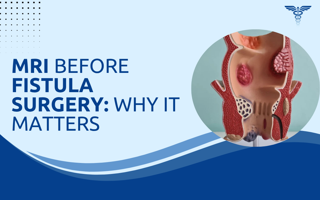
An MRI is performed before fistula surgery to provide a highly detailed 3D roadmap of the pelvic region. It is the gold-standard imaging modality for mapping hidden tracts, identifying secondary branches, and locating undrained abscesses, all of which directly determine surgical success. The scan also defines the exact relationship of the fistula to the internal and external anal sphincters, allowing the surgeon to plan a procedure that cures the disease while protecting continence. Missed branches and unrecognised abscesses are the leading causes of fistula recurrence, and MRI eliminates both.
According to Dr. Rajeev Premnath, a leading expert in fistula treatment in Bangalore, An MRI gives me information that clinical examination alone simply cannot, especially in recurrent or complex fistulas where a missed tract means certain failure.
What Does an MRI Detect Before Fistula Surgery?
A pelvic MRI captures the soft tissue anatomy of the anal region in fine detail, showing tracts, abscesses and muscle involvement with precision no clinical examination can match.
Tract mapping; The scan traces the full length of the fistula from its internal opening at the anal canal right out to the skin, including any twists or curves along the way.
Hidden branches: Secondary tracts that don’t appear during clinical examination often show up clearly on MRI, and missing even one of them is the most common reason fistulas come back.
Sphincter involvement: MRI shows precisely how much of the internal and external sphincter the tract crosses. Crucial information. The surgical approach depends entirely on this.
Abscess pockets: Undrained collections sitting next to the tract get picked up easily, which matters because operating on an active abscess without draining it first leads to poor healing.
Imaging shapes the surgical plan from start to finish. Without it, even experienced surgeons are working partly blind. For complex cases, an experienced proctologist will almost always request imaging before proceeding.
Who Needs an MRI Before Fistula Surgery?
Not every fistula requires imaging, but specific clinical situations make it essential rather than optional.
Recurrent fistulas: Anyone whose fistula has come back after previous surgery needs an MRI, because something was missed the first time and finding it without imaging is genuinely difficult.
Complex anatomy: High trans-sphincteric, supra-sphincteric and horseshoe fistulas almost always need scanning to map them properly, and these are the cases where surgical decisions matter most for continence.
Crohn’s disease: Patients with inflammatory bowel disease often develop fistulas with unusual patterns. An MRI helps tailor fistula treatment when the underlying disease changes the typical surgical approach.
Multiple openings: When there’s more than one external opening on the skin, imaging confirms whether they connect to one tract or several. Different problem entirely.
Skipping the scan in these cases is how recurrences happen. And patients pay the price with a second or third surgery. For more on this, see our guide on why fistulas come back after surgery.
Why Choose Dr. Rajeev Premnath?
Dr. Rajeev Premnath is a General and Laparoscopic Surgeon with over 20 years of experience, holding FRCS (Glasgow), FEBS and FACS qualifications along with international training in minimal access surgery from France and Singapore. He was the first surgeon in Karnataka to perform VAAFT for anal fistula, and he has extensive expertise in managing complex proctology cases.
Patients with recurrent or high fistulas often arrive after failed surgeries elsewhere. The difference here is preoperative planning. Proper imaging, precise surgical mapping and sphincter-preserving techniques mean better outcomes and a real chance of cure.
Worried your fistula might be more complex than it looks?
FAQs
Is MRI painful before fistula surgery?
No, MRI is completely painless and non-invasive, taking around 30 to 45 minutes.
Do I need contrast for fistula MRI?
Gadolinium contrast improves accuracy, particularly for active inflammation or recurrent disease.
How soon after MRI can surgery be scheduled?
Surgery is usually planned within one to two weeks of imaging for optimal results.
Can MRI replace clinical examination?
No, MRI complements but does not replace clinical and proctological assessment.